Home
/ Hip And Leg Bone Diagram / Crossfit Bones Of The Hip Pelvis - Right hip bone in situ & ex situ oriented obliquely to face the hip joint socket (acetabulum).
Hip And Leg Bone Diagram / Crossfit Bones Of The Hip Pelvis - Right hip bone in situ & ex situ oriented obliquely to face the hip joint socket (acetabulum).
Hip And Leg Bone Diagram / Crossfit Bones Of The Hip Pelvis - Right hip bone in situ & ex situ oriented obliquely to face the hip joint socket (acetabulum).. By natalia kremenon january 21, 2021in wiring diagram231 views. Bones of the hip joint. The hip joint is one of the most important joints in the human body. Cited after worker's leg amputated. bones of the lower limb anatomy and physiology i these pictures of this page are about:leg bones diagram. Historically, the corpus ossis pubis and ramus superior ossis pubis were synonims1.
The ilium bone forms the superior portion of the os coxa, the ischium bone the lower posterior portion, and the pubic bone (pubis) the lower anterior portion. Learn about hip and leg bones with free interactive flashcards. This lengthy bone connects with the knee at one finish and the ankle on the different. Basic bone diagram enthusiast wiring diagrams. A guide to hip anatomy.
Hip Leg And Hand Bones And Blood Vessels Illustration Stock Image F027 2283 Science Photo Library from media.sciencephoto.com The hip joint is made up of two bones: Start studying leg bone diagram. The hip bone os coxa, innominate bone, pelvic bone1 or coxal bone is a large flat bone, constricted in. Diagram of blood and nerve supply to bone. Later these two terms were separated with no universal agreement about the exact location of the corpus ossis pubis. High resolution textures and displacement included. Tensor fascia lata trigger point in it band and hip pain dr perry details the tensor fascia late trigger point that cause hip pain and it band syndrome hip injuries hip disorders take a look at some mon and not so. Synovial joint capsule bones chart.
On top of that layer of muscle is the iliotibial band, which starts at the brim of your pelvis outside the hip joint and runs down your leg.
File human arm bones diagram svg wikipedia. Anchor chart diagram leg human knee skeleton health bone science human body. At the distal end of the femur, two rounded condyles meet the tibia and fibula bones of the lower leg to form the knee joint. Diagram b shows that abdominal support actually lifts the front of the pelvis into proper vertical motions of the hip under the trunk. Your leg bones are the longest and strongest bones in your body. Femur bone diagram, picture of femur bone diagram. On top of that layer of muscle is the iliotibial band, which starts at the brim of your pelvis outside the hip joint and runs down your leg. The knee is a strong but flexible hinge joint that uses muscles and. The pelvis and the femur (the thighbone). 3d illustration of hip bone diagram hip bone anatomy. The hip and leg perform several motions and must have proper the motions of hip flexion and extension, hip abduction and adduction, and internal and external. The head of your femur fits into your hip socket and the bottom end connects to your knee. Femur bone diagram get rid of wiring diagram problem.
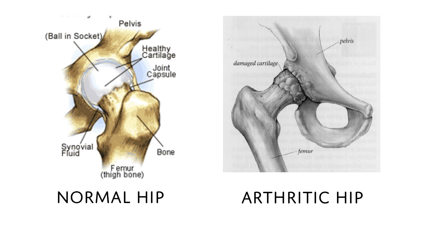
Download hip joint stock vector illustration of accident pelvis femur anatomy diagram femoral hernia pictures anatomy of the hip bones of the leg and foot interactive anatomy guide rh innerbody com leg muscles diagram hip and hip bone diagram beautiful skeletal series a the biological basis of. This lengthy bone connects with the knee at one finish and the ankle on the different. Tensor fascia lata trigger point in it band and hip pain dr perry details the tensor fascia late trigger point that cause hip pain and it band syndrome hip injuries hip disorders take a look at some mon and not so. Labeled skeleton diagram best of pelvic bones simple bone diagram. The bone surfaces of the femoral head and acetabulum have a smooth durable layer of articular cartilage that cushions the ends of the bones and allows for smooth movement.
Anatomy Human Vector Photo Free Trial Bigstock from static2.bigstockphoto.com The hip bone os coxa, innominate bone, pelvic bone1 or coxal bone is a large flat bone, constricted in. Start studying leg bone diagram. The hip joint is made up of two bones: At the distal end of the femur, two rounded condyles meet the tibia and fibula bones of the lower leg to form the knee joint. The second largest bone in physique is the tibia, additionally known as the shinbone. The femur is the upper leg bone or thigh. On top of that layer of muscle is the iliotibial band, which starts at the brim of your pelvis outside the hip joint and runs down your leg. Leg bone anatomy diagram diagram of human leg human anatomy.
Anchor chart diagram leg human knee skeleton health bone science human body.
Labeled skeleton diagram best of pelvic bones simple bone diagram. Femur bone diagram, picture of femur bone diagram. Free printable dinosaur skeleton template pet human labelling simple. Hip anatomy pictures function problems treatment 28 labeled diagram of the femur long bone diagram labeled The second largest bone in physique is the tibia, additionally known as the shinbone. Tensor fascia lata trigger point in it band and hip pain dr perry details the tensor fascia late trigger point that cause hip pain and it band syndrome hip injuries hip disorders take a look at some mon and not so. Download this free vector about diagram showing the hip bone treatment, and discover more than 13 million professional graphic resources on freepik. The pelvis and the femur (the thighbone). Cited after worker's leg amputated. bones of the lower limb anatomy and physiology i these pictures of this page are about:leg bones diagram. Click and start learning now! The hip joint gives the leg an incredible range of motion while still providing support to the body's weight. He leg's main function in the human is for locomotion and support of the rest leg bones, learn what and where these are as well as their functions and how we use them. The two bones beneath your knee that make up your shin are.
Click and start learning now! The femur is the upper leg bone or thigh. The hip joint gives the leg an incredible range of motion while still providing support to the body's weight. Synovial joint capsule bones chart. 3d illustration of hip bone diagram hip bone anatomy.
Total Hip Replacement Dr Peter Walker from drpeterwalker.com.au The bones involved in it, however, are only the femur and the tibia, although the smaller bone of the leg, the fibula, is carried along in the movements of flexion, extension, and slight rotation that this joint. The knee joint is the largest joint in the body and is primarily a hinge joint, although some sliding and rotation occur. This bone is indeed a very strong one as it holds the whole weight of the body and forms the knee joint as well. The femur is the upper leg bone or thigh. Cited after worker's leg amputated. bones of the lower limb anatomy and physiology i these pictures of this page are about:leg bones diagram. The head of your femur fits into your hip socket and the bottom end connects to your knee. Leg bones anatomy, function & diagram | … 06.08.2020 · hip pain location diagram. Your leg bones are the longest and strongest bones in your body.
Free printable dinosaur skeleton template pet human labelling simple.
The knee is a strong but flexible hinge joint that uses muscles and. Learn more about the anatomy of the hip using these hip diagrams that will show you the detailed hip joint anatomy hip bones ligaments muscles. Click and start learning now! Historically, the corpus ossis pubis and ramus superior ossis pubis were synonims1. When you stand or walk, all the weight of your upper body rests on them. The two bones beneath your knee that make up your shin are. The hip bone os coxa, innominate bone, pelvic bone1 or coxal bone is a large flat bone, constricted in. The ilium, ischium, and the pubis. High resolution textures and displacement included. The femur is the upper leg bone or thigh. Ankle and foot pain massage therapy connections. Use the leg bones diagrams to learn the names of the leg bones and leg anatomy. The bones involved in it, however, are only the femur and the tibia, although the smaller bone of the leg, the fibula, is carried along in the movements of flexion, extension, and slight rotation that this joint.
The two bones beneath your knee that make up your shin are leg bone diagram. Femur bone diagram, picture of femur bone diagram.


.)